Findings
Interpretation of nerve conduction study results
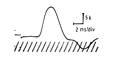
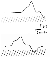
Motor Responses
The motor response originates from stimulating a nerve and recording from a muscle that it innervates. The selected muscle should possess a clearly defined motor point and minimal overlap with other nerve territories to avoid interference.
Characteristics measured include:
- Amplitude: Baseline to negative peak, expressed in millivolts
- Distal Latency: Stimulus artifact onset to baseline takeoff point, measured in milliseconds
- Waveform: Typically displays one or two initial negative peaks followed by positive deflection
Supramaximal stimulation of the nerve should ensure a maximal motor response. Proximal stimulation produces lower amplitude and longer duration responses compared to distal stimulation due to temporal dispersion of motor units.
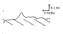
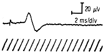
Sensory-Nerve Action Potentials (NAP)
Sensory responses are obtained by stimulating a nerve and recording directly from it or one of its branches. Recording sites must remain distant from muscles to prevent obscuration by muscle activity.
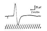
Measurement parameters include:
- Amplitude: Peak positive to peak negative deflection, expressed in microvolts
- Latency: Stimulus artifact to takeoff or negative peak
- Duration and Synchrony: Determines response characteristics based on axonal velocity dispersion
Distal Latency
Defined as the time from the stimulus affecting the nerve to the response (motor or sensory) being recorded, measured in milliseconds. This measurement enables comparison with normal reference data to assess nerve segment conductivity. Motor latency includes neuromuscular transmission delay; sensory latency does not.
Conduction Velocity
Calculated using the formula: V=d/tp-td
Where:
- d = distance in millimeters between stimulation points
- tp = proximal latency
- td = distal latency
- Result expressed in meters per second
Key principles:
- Proximal nerve segments demonstrate faster velocities
- Cold extremities slow velocity while increasing amplitude
- Anatomical entrapment points reduce velocities
- Shorter measurement segments produce less reliable calculations
- Myelin integrity primarily determines conduction velocity
- Demyelinating diseases reduce velocities below 50% of normal
- Axonal loss typically decreases velocity approximately 30% below normal
Machine Settings
Sensory Studies:
- Low frequency: 32-50 Hz
- High frequency: 1.6-2 to 3 KHz
- Sweep speed: 2 ms/division
- Sensitivity: 10-20 µV/division
Motor Studies:
- Low frequency: 1.6-2 Hz
- High frequency: 8-10 KHz
- Sweep speed: 2-5 ms/division (depending on latency/duration)
- Sensitivity: 2-10 mV/division
Distal and proximal latencies should utilize identical settings, preferably faster sweep speeds for improved takeoff identification.
Normal Values
The facility utilizes Cleveland Clinic Foundation EMG Lab normal values, categorized by patient age. Data sampling included minimum forty patients for ages 10-19 and 70+, with at least ninety patients for intermediate age groups.
Standard measurement distances:
- Median sensory: 13 cm (wrist to active electrode)
- Ulnar sensory: 11 cm
- Radial sensory: 10 cm
- Motor studies: minimum 4-6 cm (wrist to active electrodes)