The Motor Unit
Anatomy and physiology of motor unit function
Definition
An anatomical motor unit comprises an anterior horn cell, its axon, and all muscle fibers innervated by that axon and branches. Motor units vary from a few fibers in laryngeal muscles to several hundred in the gastrocnemius.
Muscle fibers from one motor unit scatter across a small muscle area, intermingled with fibers from other units.
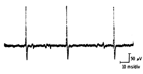
Motor Unit Action Potential: The electrical field generated by one motor unit’s muscle fibers, recorded by a nearby needle electrode.
Normal Function: Muscle fibers within one motor unit typically depolarize and repolarize synchronously.
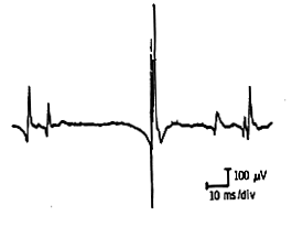
Motor Unit Potential Characteristics
Motor unit potentials are measured by:
- Amplitude: Peak-to-peak measurement
- Duration: From first baseline deflection to final return
- Phases: Number of baseline crossings plus one
- Rise time: From initial positive peak to highest negative peak
- Firing rates: Dependent on unit type and size
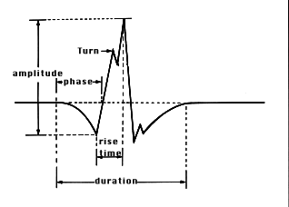
The number of phases depends largely on the synchrony of depolarization of its muscle fibers and can be affected by nerve or muscle disease.
Rise Time and Recruitment
Rise time typically measures 200-300 microseconds and depends on needle proximity to contracting fibers. Smaller units recruit early with weak effort and fire faster than larger units recruited during increased effort.
Variables Affecting Measurements
Characteristics vary with age, muscle studied, and muscle temperature. A 0.12 mm needle position shift can decrease amplitude by 50%; at 1 mm distance, amplitude decreases by approximately 90%.
When precise measurements are needed, store samples, photograph units, or use delay line triggering for detailed analysis.
Temperature Effect: Lower temperatures increase motor unit duration and amplitude.
The Needle Examination
The examination occurs in four stages: rest, mild effort, moderate effort, and full voluntary effort.
The Muscle At Rest
Insertional Activity: The muscle’s response to needle insertion consists of brief transient muscle action potentials (spikes), lasting seconds and stopping when needle movement ceases.
Abnormalities include:
- Decreased activity: Fibrosis or fat tissue replacement
- Prolonged activity: Early denervation or myotonic disorders
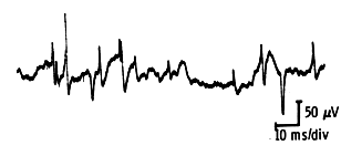
Spontaneous Activity: Persistence beyond insertion indicates activity due to end-plate noise, fibrillations, positive waves, or other spontaneous patterns.
Normal Spontaneous Activity
End-Plate Noise: Recorded near motor end-plates as either monophasic or biphasic potentials.
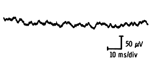
- Monophasic potentials have low amplitude and short duration, creating a “thickened baseline” appearance with “sea shell” noise or “roar” on loudspeaker
- Biphasic activity consists of irregular biphasic spikes (100-300 µv) with short duration
Examination Protocol
The resting muscle requires examination in four to five directions after needle insertion to ensure adequate sampling. Allow 0.5-1 second pause between insertions for observing spontaneous activity. When fasciculations are suspected, use 10-15 second pauses instead.
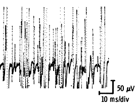
Optimal Settings for Insertional Activity:
- Oscilloscope sweep: 10 ms/division
- Amplification: 50-100 µv/division
- Low frequency filter: 32 Hz
- High frequency filter: 8000+ Hz
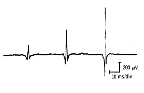
The Muscle During Voluntary Effort
Assessment occurs across three effort stages: mild, moderate, and full. Individual motor units can be studied during mild and moderate effort to measure amplitude, duration, and phases. Recruitment and firing rates are best assessed during moderate effort; interference pattern during full effort.
Mild Effort
Only a few motor units are visible at this stage—specifically the smaller units recruited first. Subjects maintain steady minimal contraction while the muscle is sampled in four to five different areas. Sample at least 20 motor units and calculate average amplitude, duration, and phase count.
Moderate Effort
Motor unit firing rates and recruitment are optimally assessed here. As muscle effort increases, firing rates accelerate and new units recruit. Units visible at this stage are larger than those appearing during mild effort.
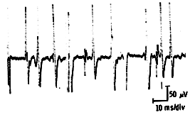
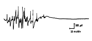
Full Effort
At maximum contraction, firing rates increase further and additional motor units recruit, making individual distinction difficult. Complete recruitment produces an interference pattern.
Motor Unit Potential Recording Settings
Motor unit potentials use the same filter settings as insertional/spontaneous activity:
- Low frequency: 16-32 Hz
- High frequency: 8000+ Hz
- Duration measurement amplification: 100-200 µv/division
- Amplitude measurement amplification: 500 µv–2000 µv/division
- Sweep speed: 5-10 ms/division
Settings vary between labs but must remain consistent within each facility for reliable measurements.